New 3D X-ray research could reshape injury treatment and rehabilitation.
Researchers in Kingston are challenging long-held beliefs about how the human shoulder moves, using advanced imaging to watch bones in motion.
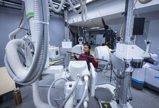
The study, led by teams at Kingston Health Sciences Centre and Queen’s University, is the first in Canada to use biplanar videoradiography, a specialized X-ray system that captures real-time joint movement. Findings were published in the Journal of Biomechanics. Lab director Mike Rainbow explains.
Working in the Skeletal Observation Laboratory at the hospital’s Hotel Dieu site, researchers combined dynamic X-ray images with CT-based 3D bone models to better understand shoulder motion during pushing and pulling tasks. Rainbow adds facilities like this are rare.
Lead author Erin Lee said the goal was to study healthy shoulders before injury occurs, helping separate cause from effect in pain-related research. Rainbow praises her work.
The team found that the shoulder blade adjusts differently depending on the type of movement, even when the arm reaches the same position. This challenges the long-standing assumption that more upward movement is always better.
Rainbow says the findings could help refine rehabilitation and surgical approaches.
Researchers say the work highlights the value of collaboration between engineers and medical teams, with potential benefits for patient care and injury prevention.
He also highlights the benefits of Queen’s University partnering with KHSC.
Researchers say the findings could lead to better treatments and help prevent shoulder injuries before they start.
Story by Alyssa Brush
 Queen's University rolls out the welcome mat for the 100th Homecoming
 Loyalist Township looking to add paid on-call firefighters during recruitment drive
 OPP reminds drivers to give cyclists at least one metre of space
 Kingston adds 10 family doctors as recruitment program reaches 43
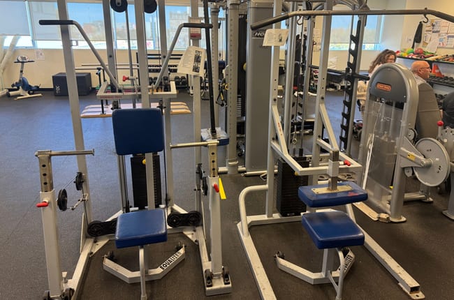 Queen's to put donated fitness equipment to use in kinesiology department